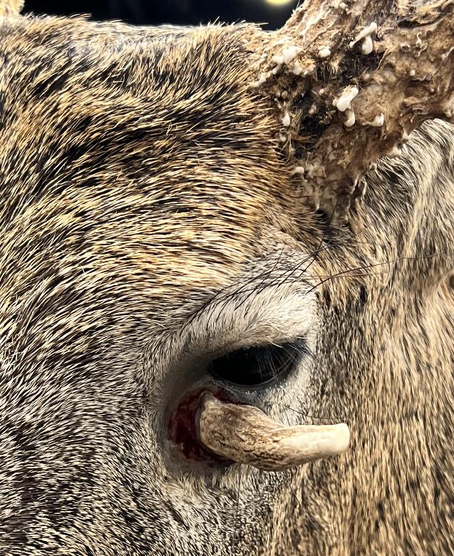
This is a very freaky — and very real — photograph of an antler sprouting from the most unusual of places.
This is a photo of a mount of the Dave Stuckey Buck from Ohio. I snapped this photo while attending the Archery Trade Show in Indianapolis last week.

In case you missed it, this buck was killed last year around Halloween by Stuckey, a renowned big-buck hunter from the Buckeye State. The buck’s rack scored 228 nontypical inches, including the bizarre 2-1/2-inch spike that was protruding out of its eye socket.
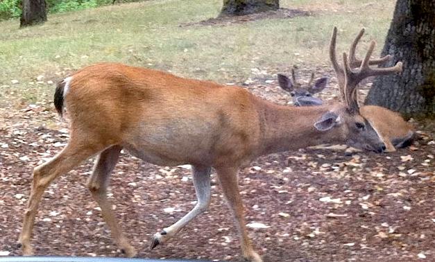
I have seen similar cases of random antlers over the years here at D&DH, but this is the largest buck (with overall rack) that I can recall. What causes such antler growth? In short, a freak happenstance of nature.
What Causes Antlers to Grow From Eye Sockets?
Perhaps the most complete scientific report on such phenomenon was reported nearly 40 years ago by renowned deer researcher Richard Goss. One of his studies, reported with R.S. Powell, was published in the Journal of Experimental Zoology in September 1985. In that report, Goss writes:
“When discs of frontal periosteum from presumptive antler sites of 6-8 month old male fawns of the fallow deer are grafted beneath the foreleg skin, they will differentiate into pedicle bones and induce small antlers in the overlying integument. These antlers shed their velvet in the fall, and in succeeding years are replaced by larger outgrowths not exceeding 7 cm in length. Periosteal transplants 1.5 cm in diameter gave rise to ectopic antlers in 100% of the grafts, while discs measuring 1.05 cm, 0.75 cm and 0.4 cm did so in only 20% of the cases. Conversely, the donor sites produced antlers in 20-23% of the cases following removal of 1.05 cm or 1.5 cm of periosteum, while 80% and 100% grew antlers after deletions of 0.75 cm and 0.4 cm discs of periosteum, respectively. Semicircular grafts of periosteum induced antler development in most cases, especially when derived from the lateral halves of the antlerogenic region on the frontal bone. These findings confirm that the histogenesis of a deer’s first pedicle and antler resides in the frontal periosteum over an area about 1.5 cm wide. They also show that leg skin is capable of antlerogenic development under the inductive influence of frontal periosteum, and that integumental wounding may enhance inductive interactions.”
In layman’s terms, Goss and Powell excised periosteum membranes from buck fawn pedicles, sutured them to other bones, and grew antlers. Fascinating stuff.
According to Medline, the periosteum is a fibrous sheath that covers bones. It contains the blood vessels and nerves that provide nourishment and sensation to the bone. The periosteum tends to be thicker in younger people. The thickness decreases when the bones are mature. The periosteum also allows bone to grow and remodel after fracture.
Final Thoughts
What about the eye socket antlers, as shown in the buck here? The thinking is similar, however, the speculation is that an injury to the periosteum layer near the eye socket triggered a similar result. It also can only be speculated that the close proximity of the eye socket to the pedicle base caused the necessary trigger for antler growth. Hence the reason why we wouldn’t see similar sporadic antler sprouts from injuries to other bone-covered periosteum layers throughout the body.


